Media resources
Completely locked-in man uses brain-computer interface to communicate
The system, developed for people with complete locked-in syndrome, allows communication over periods of months to years, in the home environment. It paves the way for new technologies for people with severe paralysis.
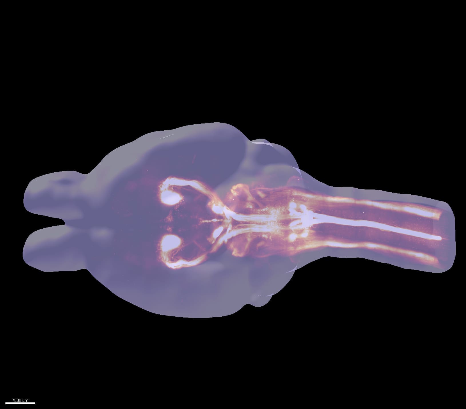
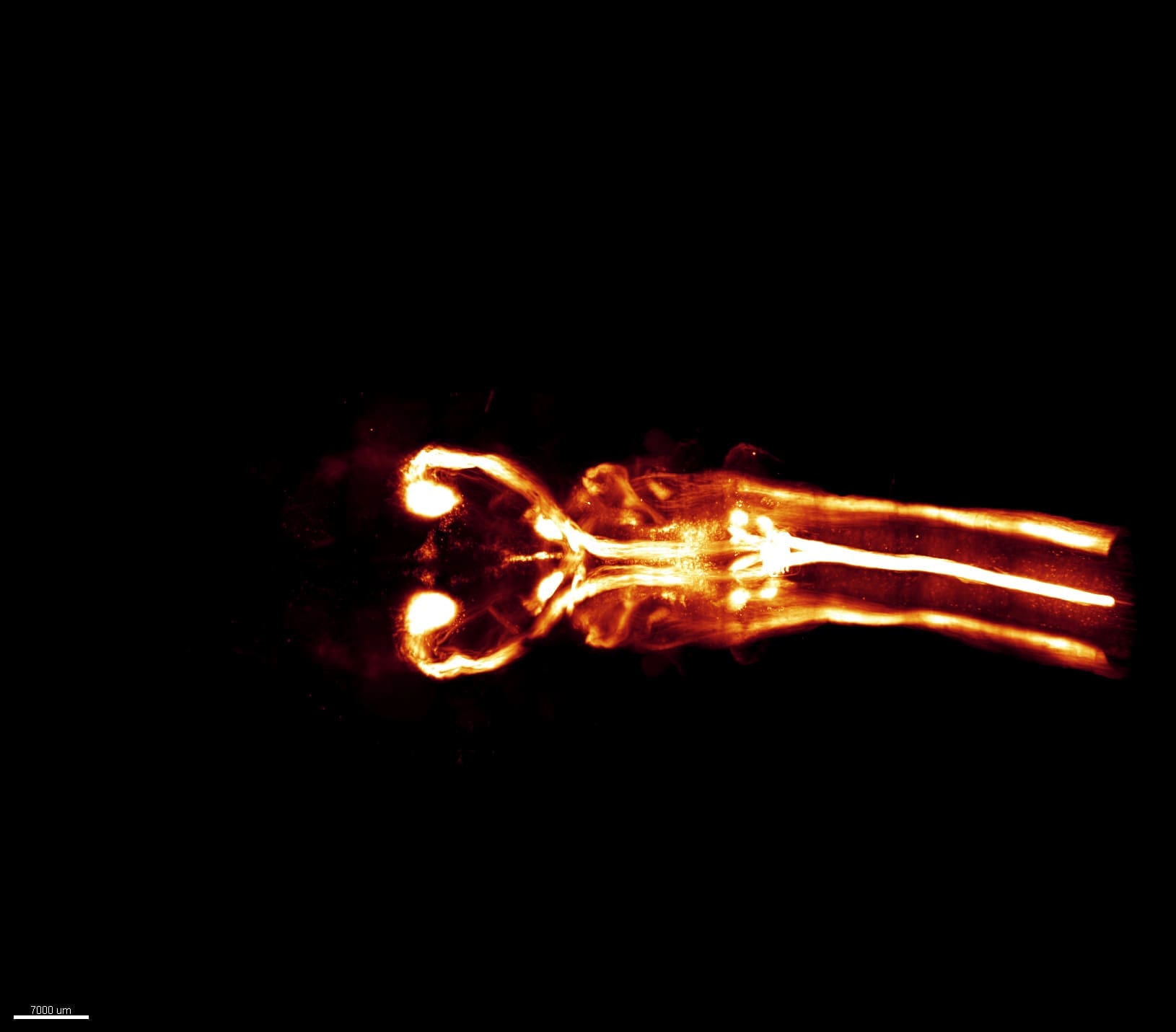
Revealing 3D anatomy with lightsheet microscopy
Developers around the world are pushing microscope technology to the extremes. Lightsheet microscopes image brain tissue down to individual neurons and offer unprecedented maps of nervous system structure and function.
Discovering new sub-cellular worlds in 3D brain samples
3D spatial transcriptomics may hold clues to future therapies for brain diseases. The technique measures gene expression from the RNA found in cells. It tells us what type of cell it is, exactly where it is, and can also indicate how a cell may interact with neighboring structures to the sides, above or below.
Image of the month: Exploring the Spiders’ Den
This image showcases a dense population of astrocytes within a sample of a human glioblastoma, recognized as the most prevalent and aggressive form of brain tumor. Originating from the biobank of HUG, this tissue sample underwent cutting-edge processing, imaging and analysis at the Wyss Center. These tumor-associated-astrocytes proliferate within the tumor, constituting a crucial element of the specific microenvironment that fosters tumor growth while evading immune detection.
At the Wyss Center, in collaboration with the Paris Brain Institute and Unige, we are pioneering the assembly of the most extensive 3D collection of human glioblastoma specimens ever compiled, utilizing our state-of-the-art lightsheet microscope. This groundbreaking collection promises to unveil crucial spatial characteristics of immune cell infiltration within glioblastomas, illuminating potential correlations with patient survival rates.
Our goal is to use this extensive dataset to understand how the tumor interacts with its microenvironment and the immune system allowing us to construct an AI-driven predictive model to develop personalized medicines, establish novel treatments and increase clinical efficacy.
Expansion microscopy: A technique to visualize the intricacies of the brain
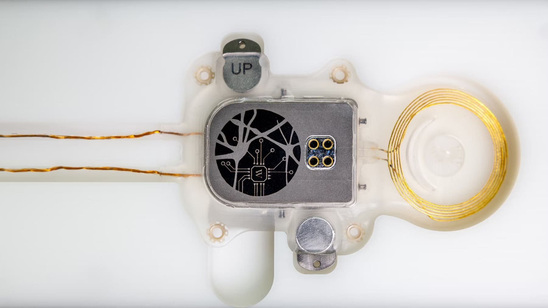
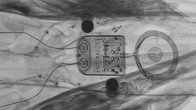
ABILITY enters preclinical trial
A preclinical trial is underway with the ABILITY brain-computer interface system.
The study, which is being carried out in sheep, is a crucial step towards development of a fully implantable device to enable applications such as communication and movement for people with paralysis.
The trial will assess the safety and feasibility of brain signal recording and wireless transfer of neural data to a wearable computer.
Where I work: Seeing the invisible