NeuroGI
Innovating technologies in neurogastroenterology to address gut-brain interaction and neurological disorders

Goal | Innovate technologies for gut-brain axis
Status | ongoing
Timeframe | 2021 – 2028
Area of Research | Neurotechnology, Neurogastroenterology, Functional Measurements
Partners | ERC, INSERM, University of Strasbourg
Lead | Michalina Gora
NeuroGI technology: From early feasibility to human concept
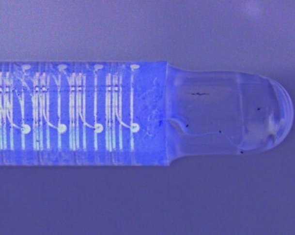
The NeuroGI technology is a minimally-invasive approach to measure signals of gastrointestinal physiology in-vivo. Initiated with the support of the European Research Council, we aimed to gain insight into signals underlying gut function in anesthetized small animals. The NeuroGI technology combines high spatiotemporal resolution recording of electrophysiology and optical coherence tomography imaging of organ morphology. By making minimally-invasive measurements of gut physiology accessible, the miniature endoscope enables longitudinal studies of this kind for the first time.
Now, our focus is to apply the know-how gained over the last 4 years to create a medical device to address the needs of patients with disorders of gut brain interaction and neurological disorders. With a core approach rooted in scientific evidence, the added support of key opinion leaders in academic and clinical neurogastroenterology will ensure the NeuroGI technology reaches clinical impact.

The gut-brain axis
Dysfunction of gut-brain communication pathways is at the core of today’s frontier in neurogastroenterology. The digestive tract is coordinated by an extensive network of neurons known as the enteric nervous system (ENS). Comprising more than 500 million neurons, the ENS controls gut motility, nutrient absorption, immune regulation and defense. Gut neurons form a wide network of cells interconnected by fibers throughout the gastrointestinal tract that directly connects to the central nervous system via multiple pathways.
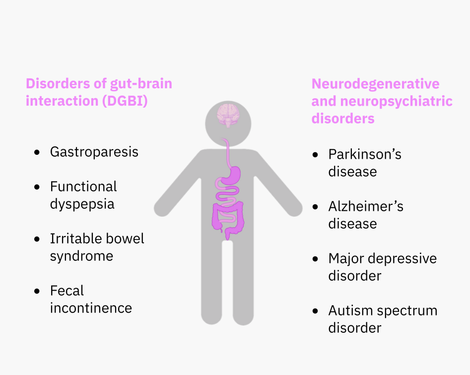
Approximately 90% of nerve fibers transmit physiological signals from the gut to the brain, thus forming the gut-brain axis. This communication is known to affect mood, appetite and memory and is implicated in disorders of gut-brain interaction and neurological disorders such as Irritable Bowel Syndrome and Parkinson’s disease.
Bridging neurobiology and engineering with advanced microscopy
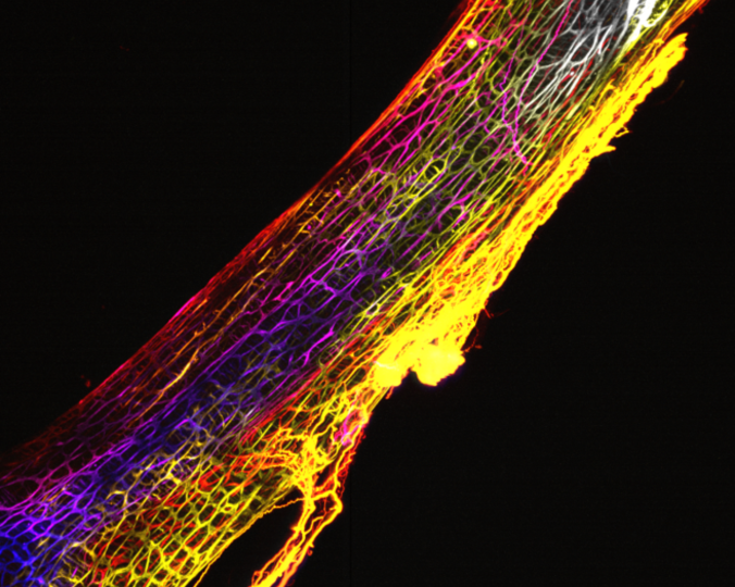
Advanced imaging techniques developed at the Wyss Center have empowered NeuroGI’s innovation by providing high resolution 3D morphological information to guide device design and engineering.
The NeuroGI team adapted the existing iDISCO protocol for optimal sample processing and lightsheet imaging of mouse and human gastrointestinal tissues. This new protocol, dubbed enteric network Gastrointestinal Lightsheet Imaging Workflow – enGLOW, enables tissue architecture observation and quantitative analyses in cubic centimeters of uninterrupted tissue and in 3 dimensions. With these capabilities, a deeper understanding of tissue morphology was applied as a neurobiology-driven guide for engineering and microfabrication of the NeuroGI mini-endoscope.
Bidirectional gut-brain communication is critical for the health of the gut, as well as the health of the brain. Disorders of gut-brain interaction, such as irritable bowel syndrome (IBS), are caused by miscommunication between these two centers and affect 40% of the population. Overall, 60% of the world population suffers from gastrointestinal symptoms.
Colonies of microbes in our gut produce over 50% of the dopamine in the body and 90% of the serotonin – both important neurotransmitters in the brain that influence feelings of pleasure and happiness. The increasing evidence that brain function is linked to gut health is leading scientists to explore the gut-brain connection in search of biomarkers and new treatment approaches for diseases including Parkinson’s disease, dementia and depression.
Despite the emerging realization of the importance of the ENS for brain and digestive health, there is no single solution to monitor the morphology and function of the ENS and its microbiota nor determine how it may change in the context of disease. Tackling the technical challenges to enable investigations of the gut-brain connection could pave the way for innovative therapies to solve major brain disorders.
Patients suffering from gastrointestinal dysfunction
Gastrointestinal symptoms affect 60% of the population. Diagnostic tools are lacking, making the field rely on self-reporting and response to medication for diagnosis. Understanding gut dysfunction is the first step towards identifying effective therapies for the brain, via the gut-brain axis.
Beachhead Market:
- Well defined unmet clinical need
- Relatively low engineering efforts
- Network of KOLs for validation
- Collection of data


