New book release: Light Sheet Microscopy
We are pleased to highlight the release of Light Sheet Microscopy, a new book in the Springer Neuromethods series that offers a comprehensive and practice-oriented guide to light-sheet fluorescence microscopy (LSFM), one of the most powerful imaging techniques for large, three-dimensional biological samples.
Widely adopted for its ability to image cleared tissues at high resolution and scale, LSFM has become an essential tool in neuroscience, developmental biology, and pathology. This book addresses both the fundamentals and the latest technical advances in the field, while remaining firmly grounded in real laboratory practice.
A structured guide from fundamentals to advanced applications
The volume is organized into three complementary sections:
- Part I introduces the principles and history of light-sheet microscopy, and key approaches to tissue clearing.
- Part II explores recent technical advances in LSFM, including improvements in resolution, field of view, and image contrast.
- Part III offers applied guides to LSFM use in diverse contexts, from imaging facilities and human brain samples to mapping immediate early genes and in vivo functional imaging in zebrafish.
Each chapter provides step-by-step detail, key notes, and expert advice designed to support reproducibility and successful implementation.
Wyss contribution: operating LSFM in a multi-user facility
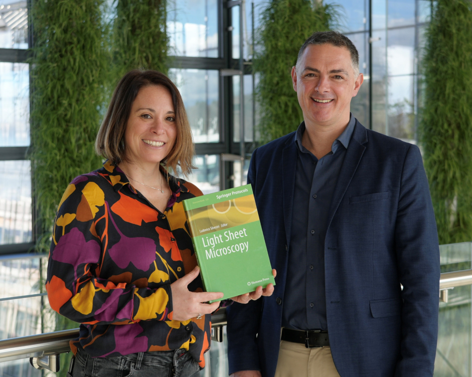
Wyss specialists, Laura Batti and Stéphane Pagès contributed a dedicated chapter entitled “Light-Sheet in a Multi-user Facility.” Their chapter focuses on the operational realities of running light-sheet microscopy platforms in shared research environments, where users may have diverse scientific questions, sample types, and levels of technical expertise.
The chapter describes the challenges inherent to providing access to LSFM at scale, including sample preparation, mounting and labeling strategies, safety considerations, system maintenance, and long-term sustainability, while outlining practical solutions developed within a multi-user imaging facility. By addressing both technical and organizational aspects, the chapter offers guidance that is directly applicable to research centers aiming to make advanced 3D imaging broadly accessible.
Enabling scalable 3D imaging for neuroscience and beyond
By combining methodological rigor with real-world facility experience, Light Sheet Microscopy serves as a valuable reference for researchers and imaging specialists seeking to adopt or optimize LSFM workflows. The contributions from Laura Batti and Stéphane Pagès highlight how thoughtful facility design and expertise can unlock the full potential of light-sheet microscopy, supporting scalable 3D imaging for neuroscience, pathology, and related fields.
Light Sheet Microscopy
This volume was edited by Ludovico Silvestri, Associate Professor at the University of Florence.
Light-Sheet Microscopy is a valuable resource for all researchers who are interested in learning more about the basic and advanced uses of LSFM.