STAR
Solutions Through Astrocyte Research
Goal | understanding the role of astrocytes in memory processes
Status | completed
Timeframe | 2022 – 2025
Area of Research | Neuro-visualization technology
Partners | Gliapharm
Lead | Prof. Andrea Volterra
Evolution | spun out as Aleos Bio

Advancing strategies to target astrocytes for the treatment and prevention of neuro-disorders
There have been few discoveries leading to drug treatments for brain diseases in recent decades and alternative avenues of investigation are urgently needed. Neurons have traditionally been the stars of brain research while the star-shaped astrocytes – which make up at least 30% of brain cells – were thought to be limited to a supporting role. There is now evidence that astrocytes are as important as neurons in assuring brain performance. In addition, astrocyte dysfunction is evident in numerous brain disorders including Alzheimer’s, Parkinson’s and Huntington’s diseases, amyotrophic lateral sclerosis, epilepsy, anxiety and depression.

Do astrocytes control memory?
Alterations in the hippocampus – the brain’s memory center – can lead to cognitive impairment and circuit malfunctions in pathologies such as Alzheimer’s and epilepsy.
Studies suggesting that astrocytes are integrated into the hippocampal circuit, and contribute to memory consolidation and performance, have resulted in growing interest in the role of astrocytes in memory.
Unlike the electrical signals of neurons, astrocytes use chemical signals – changes in their intracellular calcium (Ca2+) concentration – to communicate among themselves and with other nearby cells, including neurons.
Abnormal signaling of astrocytes in the hippocampus is thought to cause, or contribute to, cognitive impairment.
Using mini endoscopes to measure astrocyte Ca2+ signals in the brains of mice with Alzheimer’s disease, and normal mice, during learning and memory tasks, the team aims to identify how astrocytes help encode memory and how they are involved in memory alteration in Alzheimer’s disease.

Understanding the language of astrocytes
To probe how astrocytes contribute to hippocampal memory processing, the team is using live cell imaging to decode the exact mode of astrocyte communication. Some features of astrocyte Ca2+ signaling lead to the release of gliotransmitters – neuroactive molecules that are important potential targets for future therapies.
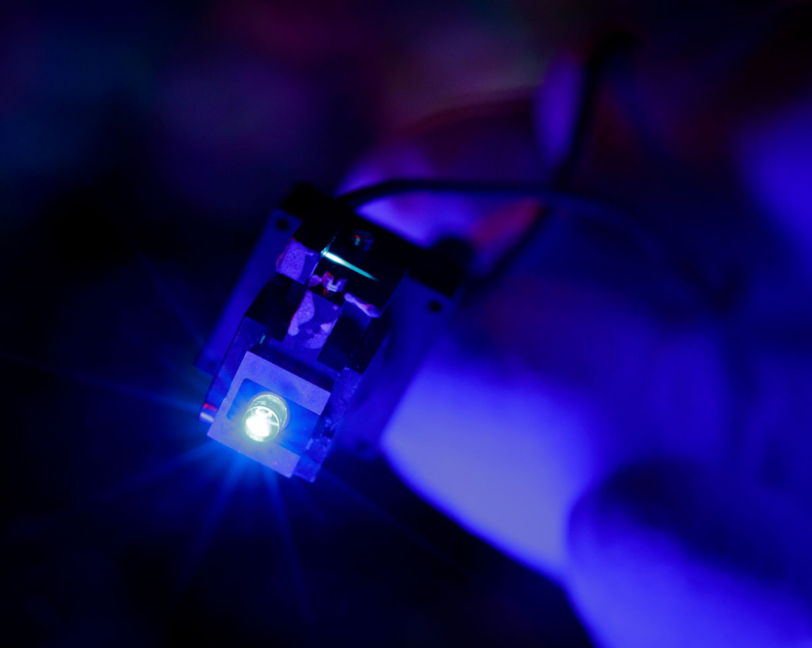
Using dynamic two-photon imaging – a technique that uses femtosecond pulsed lasers to image activity in living cells with high resolution – the team aims to identify the trigger signals coming from neuronal circuits, and the cellular mechanisms in astrocytes that lead to the release of gliotransmitters.

Tracing astrocyte connections in the brain
To further investigate the role of astrocytes in the hippocampal circuit, the team will determine if the circuit comprises connections between neurons only, as generally accepted, or also connections between astrocytes and neurons.
Using neural tracing approaches to label connections, the approach will show if astrocytes form unusual paths in the hippocampal circuit that could carry an additional layer of information. Integrated maps will reveal the position and structure of astrocyte connections as well as the associated functional and molecular information.
How astrocytes contribute to Alzheimer’s disease and epilepsy
Inflammation of the brain is a hallmark of several neurological and psychiatric conditions. Recent studies in mice and humans show that hippocampal astrocytes undergo alterations in certain genes at early disease stages, resulting in an increase in inflammatory and neurotoxic astrocytes. Cytokines – molecules involved in the immune response – are among the mediators of normal astrocyte signaling. In pathological brain conditions with inflammation, cytokine levels rise, causing dramatic changes in astrocyte-neuron interactions that contribute to cognitive alterations. To tease apart the connection between astrocyte inflammation and information processing in the brain during disease the team will assess how deletion of an astrocyte cytokine signaling pathway affects the molecular, functional and behavioral alterations that develop during Alzheimer’s disease progression and epilepsy in mice with the aim of uncovering innovative therapeutic targets.
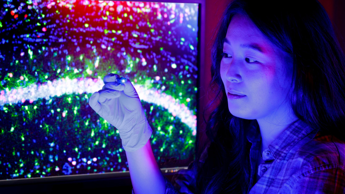
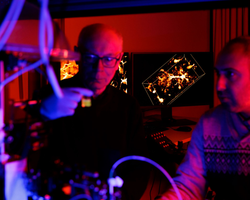
The team is using the advanced imaging pipeline at the Wyss Center to prepare, fluorescently label, image and analyze neural tissue. The state-of-the-art lightsheet microscopes, and other imaging capabilities, reveal the cellular, molecular and genetic characteristics of astrocytes within neural circuits in three-dimensions at high resolution.

The hippocampus of an Alzheimer’s mouse with astrocytes in green (GFAP labelling)

A region of the hippocampus with astrocytes labelled in green, activated neurons (c-fos-positive) in pink, and nuclei of all cells in blue (DAPI).

Confocal picture showing astrocytes (white, GFAP labelling), β-amyloid plaques (red) and nuclei of all cells (labeled in blue with DAPI). A single granule cell neuron patched with a green dye is also visible.

Astrocyte in human gbm tumor
Wyss Center researchers are supported by quality, regulatory, clinical, and entrepreneurship teams to give innovations the best opportunity of successful translation.
An existing collaboration with Gliapharm, a biotech company specializing in the research and development of novel therapies to treat and prevent neurological diseases by targeting astrocytes, will contribute to the translational objectives of the STAR project.

“If we can reveal the hidden role of astrocytes in the brain, we may unleash a secret weapon against diseases like Alzheimer’s.”

“Driven by our internationally recognized team of astrocyte experts, we are bringing together innovative ideas, cutting-edge tools and exclusive genetic models in pursuit of urgently needed treatments to fight brain disorders.”