NeuroGI
Innovating technologies to address gut-brain interaction and neurological disorders
Goal | Innovate technologies for gut-brain axis
Status | ongoing
Timeframe | 2021 – 2028
Area of Research | Neurotechnology, Neurogastroenterology, Functional Measurements
Partners | ERC, INSERM, University of Strasbourg
Lead | Michalina Gora
Healthy gut – happy brain?
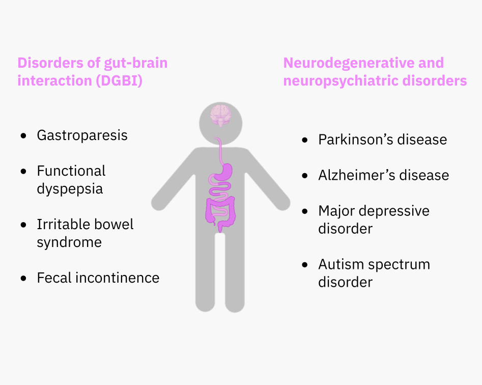
Bidirectional gut-brain communication is critical for the health of the gut, as well as the health of the brain. Disorders of gut-brain interaction, such as irritable bowel syndrome (IBS), are caused by miscommunication between these two centers and affect 40% of the population. Overall, 60% of the world population suffers from gastrointestinal symptoms.
Colonies of microbes support the healthy development of the immune and nervous systems, including the gut’s local enteric nervous system. Microbe metabolism by-products also directly affect brain functions like mood and memory. The increasing evidence of links between brain function and gut health is leading scientists to explore the gut-brain connection in search of biomarkers and new treatment approaches for diseases including Parkinson’s disease, dementia and depression.
The digestive tract is governed by a vast neural network comprising more than 500 million neurons that coordinates motility, nutrient handling, immune responses, and barrier integrity. This intrinsic neural circuitry is closely connected to the central nervous system, with the majority of neural signaling transmitting physiological information from the gut to the brain. Through this continuous dialogue, gut activity shapes processes such as mood regulation, appetite, and cognitive function.
Despite the emerging realization of the importance of the enteric nervous system (ENS) for brain and digestive health, there is no single solution to monitor the morphology and function of the ENS and its microbiota, nor determine how it may change in the context of disease. Tackling these technical challenges could pave the way for innovative therapies targeting major neurological disorders.
Research pillars
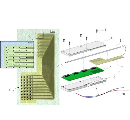
The NeuroGI mini-endoscope is opening new avenues for understanding dynamics of the gut-brain axis in disease by providing in-vivo access to gut function and morphology in small animals.
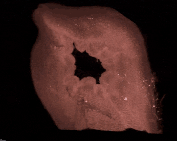
enGLOW, a state-of-the-art lightsheet microscopy workflow, enables cellular-resolution 3D imaging of labelled structures including neurons, glia, interstitial cells of Cajal and enteroendocrine cells, all of which play a crucial role in the orchestration of gut function.
By focusing on the unmet clinical need combined with cutting-edge engineering solutions, upcoming developments are aimed at diagnosing patients with disorders of gut-brain interaction in the lower gastrointestinal tract.
NeuroGI research technology
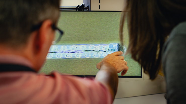
The NeuroGI technology, published in Nature Communications in February 2026, is a minimally-invasive approach to measure signals of gastrointestinal physiology in-vivo in small animals. Initiated with the support of the European Research Council, we aimed to gain insight into signals underlying gut function in anesthetized small animals. The NeuroGI technology combines high spatiotemporal resolution recording of electrophysiology and optical coherence tomography imaging of organ morphology to put the electrical measurements in the context of tissue status and motion.
Myogenic activity of the gut can be recorded in its baseline state and it can be modulated using pharmacological intervention or electrical stimulation. By benchmarking against gold standard in collaboration with prof. Nick Spencer, we confirmed that orchestration by the ENS is critical for coordinated functions observed in-vivo. By making minimally-invasive measurements of gut physiology accessible, the miniature endoscope enables longitudinal studies of this kind for the first time.
Function in the context of morphology to bridge neurobiology and engineering
Advanced imaging techniques developed by the ALICE platform at the Wyss Center have empowered NeuroGI’s innovation by providing high resolution 3D morphological information to guide device design and engineering.
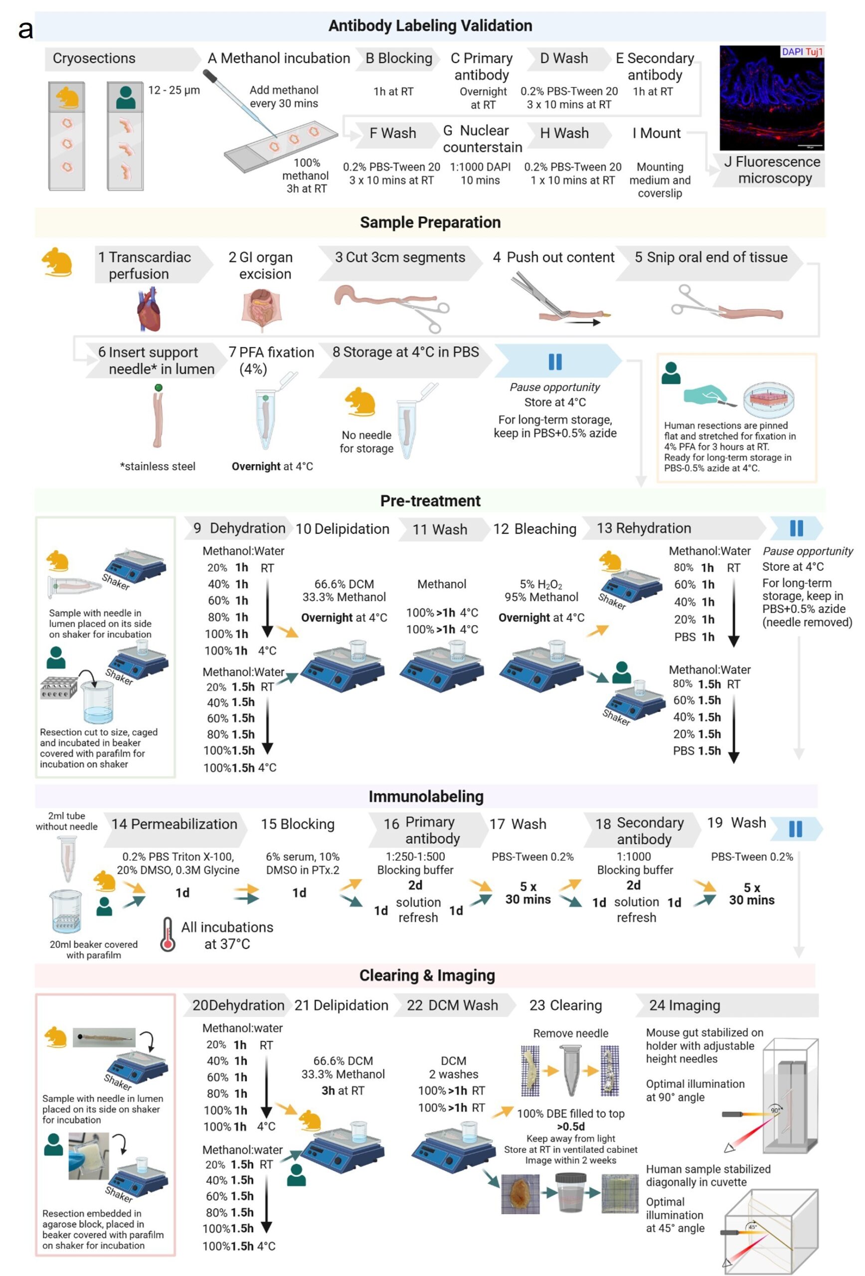
The NeuroGI team adapted the existing iDISCO protocol for optimal sample processing and lightsheet imaging of mouse and human gastrointestinal tissues. This new protocol, dubbed enteric network Gastrointestinal Lightsheet Optical Workflow – enGLOW, enables tissue architecture observation and quantitative analyses in cubic centimeters of uninterrupted tissue and in 3 dimensions. With these capabilities, a deeper understanding of tissue morphology was applied as a neurobiology-driven guide for engineering and microfabrication of the NeuroGI mini-endoscope.
enGLOW, published in Communications Biology in February 2026, offers guidance starting from gut sample collection so that researchers can obtain images and quantify morphology parameters. The NeuroGI team is currently applying it to advance neurobiology understanding of ENS remodeling and neuroinflammation in multiple disorders, including body-first Parkinson’s disease in collaboration with Prof. Nathalie Van Den Berge (Aarhus University) and alcohol addiction in collaboration with Prof. Laura Harsan (University of Strasbourg).
Towards translation for clinical impact
With a core approach rooted in scientific evidence obtained from animal models, we work closely with key opinion leaders in academic and clinical neurogastroenterology to identify and address the field’s unmet clinical needs. The NeuroGI project’s next step is translating our intraluminal approach of electrophysiology-based assessment of gut function to the human gastrointestinal tract. Thus, relying on the know-how gained since the start of the project, we are advancing towards the first prototype of a human-grade medical device. This solution will aim to improve diagnosis of disorders of gut-brain interaction, such as irritable bowel syndrome, and identify risk factors for neurological disorders with early-onset gut dysfunction, such as Parkinson’s disease.
Interdisciplinary team bringing together neuroscience and gastroenterology
Minimally invasive intraluminal approach to preserve physiological function
New paradigm for diagnosis and treatment of the gut and the brain
Publications

Michalina Gora
Group Leader
PhD

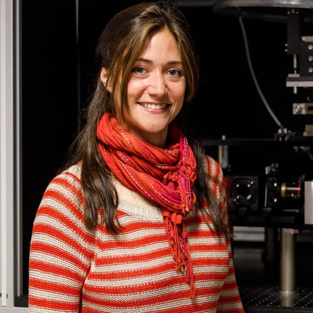
Arielle Planchette
Research Scientist
PhD
Aleksander Sobolewski
Staff Neuroscientist
PhD

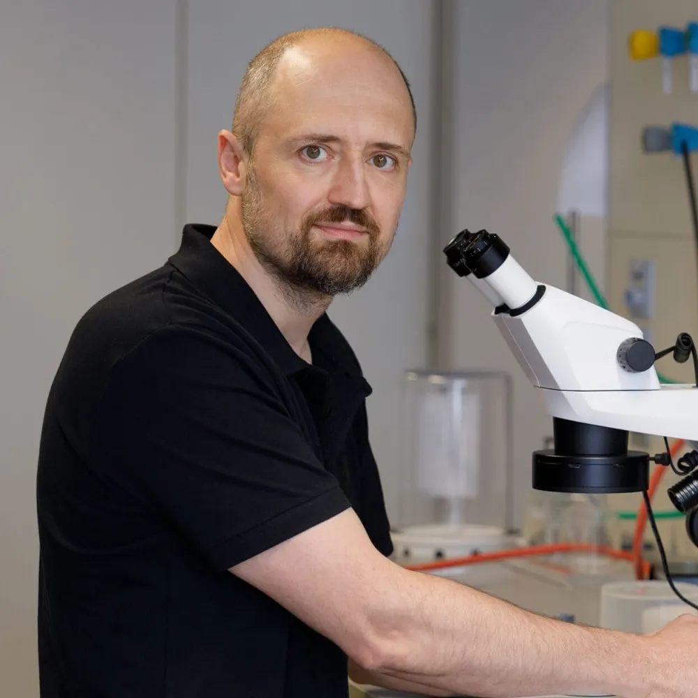
Karol Wojcicki
Mechanical Engineer / Lab Technical Specialist
BSc
Yoseline Cabara
Photonic Systems Engineer
PhD

Shenandoah Montamat
Senior Neurotechnology Transfer Manager
MSc

Etienne de Montalivet
Machine Learning Scientist

Karissa Humailo
Intern

